|
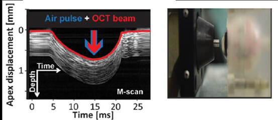
by Alejandra Consejo, Postdoc Researcher & Assistant Professor, Polish Academy of Sciences IMCUSTOMEYE will join Optical Coherence Tomography (OCT) with dynamic imaging of the cornea to assess its biomechanical properties and bring this technology into the clinic with portable instrumentation. OCT is a non-invasive imaging test. ‘Optical’ refers to light. ‘Coherence’ refers to a property of light, and ‘Tomography’ is imaging by sections (slides). OCT uses low-coherence light to capture micrometer-resolution, two- and three-dimensional images from within optical scattering media (e.g., biological tissue). Optical coherence tomography is one of a class of optical tomographic techniques. Commercially available OCT systems are employed in diverse applications, including art conservation and diagnostic medicine, notably in ophthalmology. The advent of OCT imaging has changed the way ophthalmologists image the ocular surface and anterior segment of the eye. Its ability to obtain a dynamic, high, and ultra-high resolution, cross-sectional images of the ocular surface, and anterior segment in a non-invasive and rapid manner allows for ease of use. The Physical Optics and Biophotonics Group at the Polish Academy of Sciences, lead by Prof. Maciej Wojtkowski, is a pioneer in developing new, top-class imaging systems and specialized in Optical Coherence Tomography technology. Figures 1 and 2 are examples of OCT developments performed by prof. Wojtkowski’s team and collaborators. Figure 1. Three-dimensional cutaway view of the human cornea acquired in vivo. Acquired from ‘In vivo imaging of the human cornea with high-speed and high-resolution Fourier-domain full-field optical coherence tomography’ (2020). This research work was developed by Prof. Wojtkowski’s team, and it is available in full here. Figure 2. Figure adapted from ‘Assessment of the influence of viscoelasticity of cornea in animal ex vivo model using air-puff optical coherence tomography and corneal hysteresis’ (2019), co-authored by Dr. Karnowski and Prof. Wojtkowski. Available in full here. This work uses enucleated porcine corneas to demonstrate the usefulness of imaging the cornea using OCT, while mechanically stimulating it with an air pulse, to gain information on corneal viscoelasticity.
1 Comment
11/20/2022 11:36:09 pm
Optical Coherence Tomography (or OCT) is a non-invasive imaging technique used to diagnose or monitor various eye conditions. The technology has been around for many years, but has recently become much more popular with the boom of smartphones and cellular data.
Reply
Leave a Reply. |
Proudly powered by Weebly

