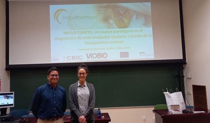
 The Science and Technology Week organized by CSIC is the most important scientific outreach initiative in Spain. It is organized every year over two weeks with the objective of bringing science closer to the public. The activity carried out by IMCUSTOMEYE was the conference “IMCUSTOMEYE: A new paradigm in the diagnosis of ocular diseases through corneal biomechanics” by Judith Birkenfeld (PhD) and Fernando Zvietcovich (PhD) on November 2022 at the Institute of Optics CSIC (Madrid, Spain). 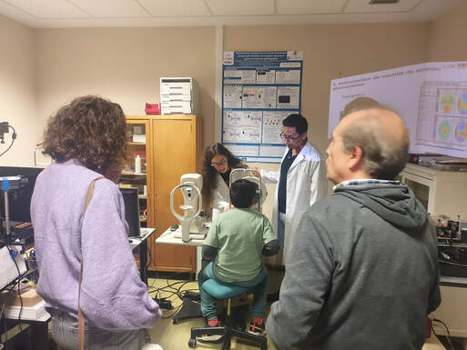 We also held an open doors day to bring the public into direct contact with the project. During this day IMCUSTOMEYE researchers organized two visits to our laboratories at the Institute of Optics (CSIC, Spain). The attendees included people of a wide range of ages, from older people to college students and children. The attendees followed the steps that a patient would follow in an IMCUSTOMEYE study at the VioBio Lab, showing them different devices like an Optical Coherence Tomography (OCT) system. The lab tours were designed to show also different dissemination tools developed throughout the project like the latest IMCUSTOMEYE video and flyer.
 The mechanical properties of biological tissues play an essential role in maintaining the structural stability and adequate function of organs in the human body. A considerable amount of recent research effort has been focused on the development of measurement techniques for the quantification of biomechanics in ocular tissues, particularly in the cornea. Corneal biomechanics contribute significantly to the maintaining of adequate corneal shape and function, therefore, abnormal biomechanical properties can be an indicator of corneal pathologies or structural deterioration such as keratoconus.
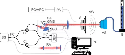
In this research, conducted by University of Galway and the Institute of Optics CSIC, we present a co-axial acoustic-based optical coherence vibrometry probe (CoA-OCV) for vibro-acoustic resonance quantification in biological tissues. Sample vibrations were stimulated via a loudspeaker, and pre-compensation was used to calibrate the acoustic spectrum. Sample vibrations were measured via swept-source optical coherence tomography (OCT). Resonance frequencies of corneal phantoms were measured at varying intraocular pressures (IOP), and dependencies on Young´s Modulus (E), phantom thickness and IOP were observed. The results of the current study demonstrate the feasibility of CoA-OCV for use in future OCT-V studies. Full reference: R. McAuley, A. Nolan, A. Curatolo et al. Co-axial acoustic-based optical coherence vibrometry probe for the quantification of resonance frequency modes in ocular tissue. Scientific Reports 12, 18834 (2022). Link to the article here 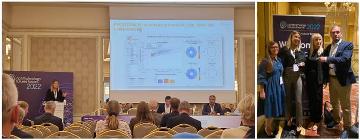 We are proud to announce that IMCUSTOMEYE was the winner of the Shark Tank session at the Ophthalmology Futures Forums that took place in Milan the past September 15th. Lisa Kleintjens, from IROC Science to Innovation AG, presented IMCUSTOMEYE technologies for corneal biomechanical mapping offering clinicians a clear insight into the biomechanical state of their patients’ eyes so that they can screen, diagnose, and treat their patients with more accuracy and safety.
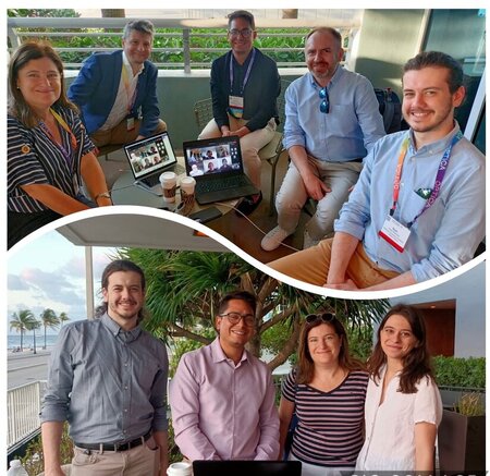 One more year IMCUSTOMEYE partners have presented new project results in several international conferences, including Optica Biophotonics Congress at Fort Lauderdale (Florida, USA) and ARVO 2022 Annual Meeting at Denver (Colorado, USA).
The Biophotonics Congress is organized by Optica (formerly OSA), the leading society dedicated to promoting the generation, application and dissemination of knowledge in the field, and, this year, was focused on biomedical optics to advance application of medical science to clinical practice. Here are IMCUSTOMEYE contributions: - Ryan McAuley, Andrew Nolan, Andrea Curatolo, Sergey Alexandrov, Fernando Zvietcovich, Alejandra Varea, Susana Marcos, Judith Birkenfeld, Martin Leahy. "Hysteresis in Vibrational Resonance Modes of Model Corneas Measured With Optical Coherence Tomography Vibrography Utilizing a Co-Axial Acoustic Stimulation Technique and Pre-Compensation". Optica Biophotonics Congress: Biomedical Optics (2022) - Karol Karnowski, Jadwiga Milkiewicz, Angela Pachacz, Andrea Curatolo et al. "Simultaneous Multi-Spot OCT Measurements of Air Induced Corneal Deformations". Optica Biophotonics Congress: Biomedical Optics (2022) - Fernando Zvietcovich, Achuth Nair, Manmohan Singh, Salavat Aglyamov, Michael Twa, Kirill Larin. "In-Vivo Assessment of Corneal Biomechanics Under Localized Cross-Linking Treatment Using Wave-Based Optical Coherence Elastography". Optica Biophotonics Congress: Biomedical Optics (2022) The ARVO congress takes place every year and plays a key role for research in vision and ophthalmology since it is the largest gathering of researchers in the field. This year we presented results on cross-meridian air-puff corneal deformation OCT and wave-based optical coherence elastography (OCE) for keratoconus detection: - Fernando Zvietcovich; Judith Birkenfeld; Alejandra Varea; Ana Maria Gonzalez; Andrea Curatolo; Susana Marcos. "Multi-meridian wave-based corneal optical coherence elastography in normal and keratoconic patients". Investigative Ophthalmology & Visual Science June 2022, Vol.63, 2380 – A0183. - Judith Sophie Birkenfeld; Alejandra Varea; Andrea Curatolo; Ashkan Eliasy; Ana Maria Gonzalez; Eduardo Martinez-Enriquez; Ahmed Abass; Bernardo Lopes; Ahmed Elsheikh; Jesus Merayo-Lloves; Susana Marcos. "Corneal biomechanical parameters in healthy and early-stage keratoconus eyes from cross-meridian air-puff deformation OCT". Investigative Ophthalmology & Visual Science June 2022, Vol.63, 2394 – A0197 In the past weeks, reserachers from CSIC VioBio Lab led by prof. Susana Marcos and from University of Liverpool Biomechanical Engineering Group led by Prof. Ahmed Elsheikh have presented the progress of the IMCUSTOMEYE project in two European congresses of great impact within the optics and ophthalmology field. The Spanish Optical Society (SEDOPTICA) triennial meeting (RNO) took place online this year. An average of 200 professionals participate in each RNO congress where the latest scientific and technological advances in the field are presented. This year’s RNO is organized by SEDOPTICA’s Women, Optics and Photonics Area. Here is IMCUSTOMEYE contribution to the congress: - Birkenfeld, J., Abass, A., Alexandrov, S., Bronte-Ciriza, D., Curatolo, A., Eliasy, A., ... & Marcos, S. Applications of the ImTOPScanner for the investigation of ocular biomechanical parameters, XIII RNO (2021).  CXL Experts' Meeting is designed to bring delegates up-to-speed on the latest best-practices in corneal cross-linking for the treatment of ectasias and keratitis. Reserachers from the University of Liverpool presented several contributios at the meeting:
- Ashkan Eliasy, Prema Padmanabhan, Bernardo T Lopes, Haixia Zhang, Ahmed Abass, Ahmed Elsheikh. Cone Shape Change with Keratoconus Progression, CXL Experts' Meeting 2021 - Bernardo T Lopes, Prema Padmanabhan, Ashkan Eliasy, Haixia Zhang, Ahmed Abass, Ahmed Elsheikh. Corneal biomechanical deterioration with keratoconus progression assessed by the Stress-Strain Index, CXL Experts' Meeting 2021 - Ahmed Elsheikh, Bernardo T Lopes, Ashkan Eliasy, Haixia Zhang, Ahmed Abass, Prema Padmanabhan. Regional variation of corneal stiffness with Keratoconus progression assessed by the Stress-Strain Maps, CXL Experts' Meeting 2021 - Ahmed Abass, Bernardo T Lopes, Ashkan Eliasy, Ahmed Elsheikh. Estimated Corneal Remodelling in Response to Intracorneal Ring Segments, CXL Experts' Meeting 2021 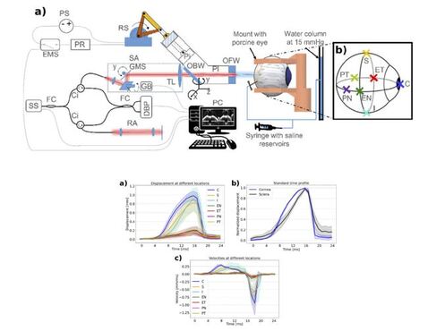 Scleral biomechanics play an important role in the progression of myopia (near-sightedness), but it is challenging to measure it non-invasively. In our recent publication we introduce air-puff deformation imaging as a method to estimate the biomechanical properties of the sclera of an intact eye globe. The sclera was excited via an air-pulse at several globe locations, and the deformation was imaged using optical coherence tomography (OCT). The maximum apex deformation, the deformation velocity, and the arc-length during deformation was investigated. The elastic modulus of the sclera was then estimated using an inverse optimization technique based on finite element modeling.
Full reference: David Bronte-Ciriza, Judith S. Birkenfeld, Andrés de la Hoz, Andrea Curatolo, James A. Germann, Lupe Villegas, Alejandra Varea, Eduardo Martínez-Enríquez, and Susana Marcos, “Estimation of scleral mechanical properties from air-puff optical coherence tomography,” Biomed. Opt. Express 12, 6341-6359 (2021) Link to the article here 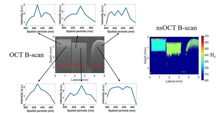 Optical coherence tomography (OCT) is a rapidly evolving technology with a broad range of applications, including biomedical imaging and diagnosis. Conventional intensity-based OCT provides depth-resolved imaging with a typical resolution and sensitivity to structural alterations of about 5–10 microns. It would be desirable for functional biological imaging to detect smaller features in tissues due to the nature of pathological processes. In this article, researchers from the National University of Ireland Galway, perform the analysis of the spatial frequency content of the OCT signal based on scattering theory. We demonstrate that the OCT signal, even at limited spectral bandwidth, contains information about high spatial frequencies present in the object which relates to the small, sub-wavelength size structures. Presented results provide a theoretical and experimental basis to substantially improve the sensitivity of OCT to structural alterations at clinically relevant depths.
Full reference: Alexandrov, S., Arangath, A., Zhou, Y. et al. "Accessing depth-resolved high spatial frequency content from the optical coherence tomography signal". Sci Rep 11, 17123 (2021). https://doi.org/10.1038/s41598-021-96619-7 Full article here  In the past months, Imcustomeye partners have presented multiple results of the project on corneal biomechanics at several international congresses including ARVO 2021 Annual Meeting, OSA Biophotonics Congress 2021, SPIE Conference "Optical Coherence Tomography and Coherence Domain Optical Methods in Biomedicine" and European Conferences on Biomedical Optics. ARVO is the largest and most respected eye research organization in the world, with 11,000 members worldwide, and its congress takes place every year playing a key role for research in vision and ophthalmology. This year Judith Birkenfeld's poster on first results with the newest device ImTOPScanner was designated Hot Topic by the organization. Here are the Imcustomeye contributions: - Judith S. Birkenfeld; Andrea Curatolo; Ashkan Eliasy et al. “Improved detection of corneal deformation asymmetries in keratoconus patients using multi-meridian deformation imaging" Investigative Ophthalmology & Visual Science Vol.62, 2031 (2021) - Lupe Villegas; James Germann; Susana Marcos. "Biomechanical effects of Scleral crosslinking using Rose Bengal/Green-light and Rivoflavin/UVA". Investigative Ophthalmology & Visual Science Vol.62, 2281 (2021) - Ashkan Eliasy; Bernardo Teixeira Lopes; Ahmed Abass et al. "Evaluation of Stress-Strain Index (SSI2) in Healthy and Keratoconic corneas". Investigative Ophthalmology & Visual Science Vol.62, 777 (2021) - Bernardo T Lopes; Ahmed Abass; Ashkan Eliasy; Haixia Zhang; Ahmed Elsheikh. "A New Method to Describe Cone Shape in Keratoconic Corneas". Investigative Ophthalmology & Visual Science Vol.62, 2025 (2021) OPTICA, formerly OSA, and SPIE are international professional societies working to advance optics and photonics. Imcustomeye has contributed with the following talks at conferences organized by these societies: - Andrea Curatolo, Angela Pachacz, Jedrzej Solarski et al. "Corrections of motion artifacts in dynamic low-cost, swept-source optical coherence tomography" in Biophotonics Congress 2021. OSA Technical Digest (Optical Society of America), paper DTh1A.4 (2021) - Karol Karnowski, Jedrzej Solarski, Andrea Curatolo et al. "Corrections of axial shift artifact in dynamic swept-source optical coherence tomography" in SPIE BiOS 2021. Optical Coherence Tomography and Coherence Domain Optical Methods in Biomedicine XXV, Proceedings Vol. 11630 (2021). - Judith S. Birkenfeld, David Bronte-Ciriza, Andrés de la Hoz et al. "Proof-of-concept method to estimate scleral mechanical properties from air-puff Optical Coherence Tomography". European Conferences on Biomedical Optics 2021.  Moorfields Eye Hospital, clinical partner of the IMCUSTOMEYE consortium, has published a post about the IMCUSTOMEYE project on their intranet and newsletter. The post introduce the project and talks about the Moorfields’ role testing patients with keratoconus, cataract and glaucoma to validate the new devices’ ability to identify corneal disease and measure eye pressure accurately.
Moorfields Eye Hospital is the leading provider of eye health services in the UK and a world-class centre of excellence for ophthalmic research and education. The main focus of their 2300 staff members is the treatment and care of NHS patients with a wide range of eye problems. The volume and variety of conditions treated by Moorfields clinicians means that they have a unique range of skills and knowledge. The next step in the project for Moorfields will be to set up the study mentioned before and then begin recruiting patients from clinics to take part in the testing. You can read the post here 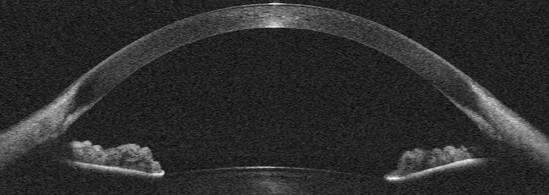 The University of Liverpool, member of the IMCUSTOMEYE consortium, has published a press release on the project in its research section. The article highlights the role of the University’s Biomechanical Engineering Group in the development of new imaging techniques to diagnose and treat corneal diseases and glaucoma in a personalised manner.
Professor Ahmed Elsheikh, head of the research group, explains that “despite the fact that these treatments, and the conditions they help with, interact or interfere mechanically with the cornea, there is currently no method to accurately measure corneal biomechanical properties in vivo. Such a method would be essential for customising these treatments for individual patients’ needs and ultimately improving their clinical outcomes. The IMCUSTOMEYE project is intended to address this need,” Check out the full press release here IMCUSTOMEYE landmark paper on first multi-meridian corneal imaging device PUBLISHED IN BOE10/16/2020
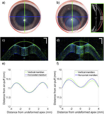 An EU Horizon 2020 IMCUSTOMEYE project landmark paper has just been published in Biomedical Optics Express. It is the result of a collaboration between CSIC, University of Liverpool, and the Polish Academy of Science. It presents the first multi-meridian corneal imaging device, based on a swept-source optical coherence tomography system with high transverse scanning frame rates, to measure a promising early keratoconus biomarker based on air-puff-induced deformation asymmetry. The system will serve both as the basis for determining the minimum viable specifications for a keratoconus screening device based on a deformation asymmetry biomarker, and as a platform to estimate patient-specific corneal biomechanical properties, that can be fed in corneal surgery planning simulators. Currently the system is employed in a pilot clinical study with Keratoconus and Lasik-candidate subjects in collaboration with Fernandez Vega Ophthalmological Institute.
Full reference: Andrea Curatolo, Judith S. Birkenfeld, Eduardo Martinez-Enriquez, James A. Germann, Geethika Muralidharan, Jesús Palací, Daniel Pascual, Ashkan Eliasy, Ahmed Abass, Jędrzej Solarski, Karol Karnowski, Maciej Wojtkowski, Ahmed Elsheikh, and Susana Marcos, "Multi-meridian corneal imaging of air-puff induced deformation for improved detection of biomechanical abnormalities," Biomed. Opt. Express 11, 6337-6355 (2020) Full article here 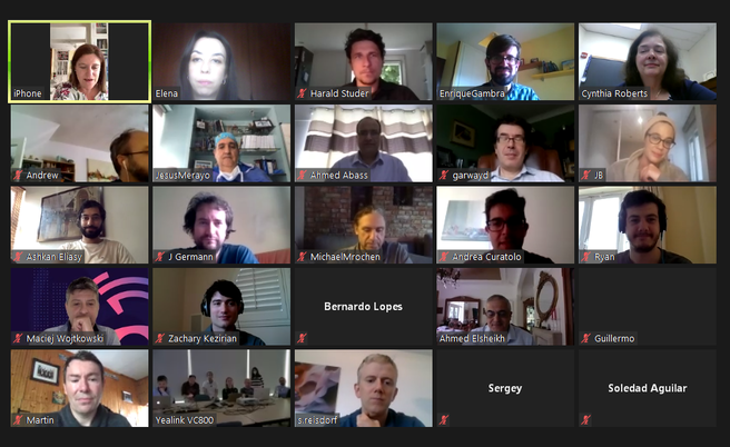 The past 15th and 16th of June took place Imcustomeye Annual Review Meeting. The event was hosted by VioBio Lab (CSIC) and the Institute of Physical Chemistry (Polish Academy of Sciences) and, due to this year exceptional circumstances, the meeting was virtual and took place via Zoom.
All partners, including the PIs, attended the two days meeting, gaving an update on the research, IP and outreach progress of the project during this past year. Professor Cynthia Roberts (Department of Biomedical Engineering, Ohio State University), member of the Advisory Board, was invited to the meeting where she gave a lecture on "The importance of corneal biomechanics as a diagnostic marker". The event also included bilateral discussions on specifics of the project between designated partners, a steering committee session and a debate on the impact of COVID-19 on market of medical devices and clinical studies. 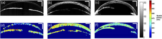 Optical coherence tomography (OCT) is a non-invasive depth resolved optical imaging modality, that enables high resolution, cross-sectional imaging in biological tissues and materials at clinically relevant depths. Though OCT offers high resolution imaging, the best ultra-high-resolution OCT systems are limited to imaging structural changes with a resolution of one micron on a single B-scan within very limited depth. Nanosensitive OCT (nsOCT) is a recently developed technique that is capable of providing enhanced sensitivity of OCT to structural changes. Improving the sensitivity of OCT to detect structural changes at the nanoscale level, to a depth typical for conventional OCT, could potentially improve the diagnostic capability of OCT in medical applications. Researchers from the National University of Ireland Galway demonstrates in this paper, published by Biomedical Optics Express, the capability of nsOCT to detect structural changes deep in the rat cornea following superficial corneal injury. Full reference: C. Lal, S. Alexandrov, S. Rani, Y. Zhou, T. Ritter, M. Leahy, “Nanosensitive optical coherence tomography to assess wound healing within the cornea”. Biomed. Opt. Express 11 (7) 3407-3422 (2020) https://doi.org/10.1364/BOE.389342 Full article here 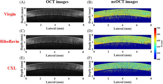 Corneal cross‐linking (CXL) using ultraviolet‐A (UVA) irradiation with a riboflavin photosensitizer has grown from an interesting concept to a practical clinical treatment for corneal ectatic diseases globally, such as keratoconus. This research led by NUI Galway reports the application of oversampling nanosensitive OCT for probing the corneal structural alterations. The results indicate that the spatial period increases slightly after 30 minutes riboflavin instillation but decreases significantly after 30 minutes UVA irradiation following the Dresden protocol. The proposed noninvasive method can be implemented using existing OCT systems, without any additional components, for detecting nanoscale changes with the potential to assist diagnostic assessment during CXL treatment, and possibly to be a real‐time monitoring tool in clinics.
Full reference: Y. Zhou, S. Alexandrov, A. Nolan, N. Das, R. Dey and M. Leahy, “Non‐invasive detection of nanoscale structural changes in cornea associated with corneal cross‐linking treatment”. Journal of Biophotonics 13 (6) e201960234 (2020) https://doi.org/10.1002/jbio.201960234 Full article here  First press release on Imcustomeye project was published by Consejo Superior de Investigaciones Científicas (CSIC) this May. The press release seeks to introduce the project to the general public, including a description of the consortium of academic experts, ophthalmic clinics and companies, the objectives of the project and an overview on corneal biomechanics. The article also includes quotes from interviews with PI Prof. Susana Marcos and postdoctoral researchers from VioBio Lab Judith Birkenfeld and Andrea Curatolo.
Link to the press release here 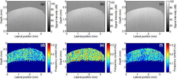 Most of the fundamental pathological processes in living tissues exhibit changes at the nanoscale. Noninvasive, label-free detection of structural changes in biological samples pose a significant challenge to both researchers and healthcare professionals. Modern nanoscopy largely requires labeling, is limited to superficial 2D imaging, and is generally not suitable for in vivo applications. In this article published in Applied Physics Letters, we presented a method, spatial frequency domain correlation mapping optical coherence tomography (sf-cmOCT), developed at NUI Galway, for detection of depth resolved nanoscale structural changes noninvasively. Our approach is based on detection and correlation of the depth resolved spectra of axial spatial frequencies of the object which are extremely sensitive to structural alterations. These experimental results demonstrate possibilities for the study of nanoscale structural changes, without the need for biomarkers or labels. Thus, sf-cmOCT offers exciting and far-reaching opportunities for early disease diagnosis and treatment response monitoring, as well as a myriad of applications for researchers. Full reference: Sergey Alexandrov, Paul M. McNamara, Nandan Das, Yi Zhou, Gillian Lynch, Josh Hogan, and Martin Leahy. Spatial frequency domain correlation mapping optical coherence tomography for nanoscale structural characterization. Applied Physics Letters 115:12 (2019). Full article here 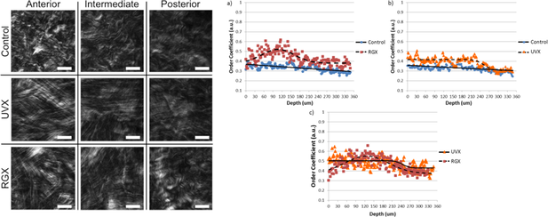 One option for treating keratoconus, a degenerative disease that results in a weakened cornea and vision loss, is cross-linking. Cross-linking strengthens the overall structure of the cornea by promoting the creation of covalent bonds between neighboring collagen fibrils or between fibrils and the surrounding extracellular matrix. To measure how cross-linking affects the cornea at different depths inside the stroma, three different corneal treatments were performed on rabbit animal models: no treatment, Riboflavin instillation/Ultraviolet irradiation, and Rose Bengal instillation/Green light irradiation. Rabbits were sacrificed one and two months after treatment. Images of corneal lamellae were taken with a second harmonic generation laser scanning microscopic developed at VioBio Lab (CSIC) and were analyzed with a MATLAB script to measure the different directions of the collagen fibers. The collagen fibers in the cross-linked corneas had more uniform fiber orientations than in untreated corneas through a depth of 300 microns. The amount of second harmonic generation is also greater in cross-linked corneas than in control corneas. Both the organization and second harmonic generation signal increased between one month and two months after treatment. This research was a collaboration between CSIC, the University of Valladolid, and the Wellman Center for Photomedicine.
Full reference: James A. Germann, Eduardo Martínez-Enríquez, M. Carmen Martínez-García, Irene E. Kochevar, Susana Marcos; Corneal Collagen Ordering After In Vivo Rose Bengal and Riboflavin Cross-Linking. Invest. Ophthalmol. Vis. Sci. 61(3):28 (2020). Full article here |
 RSS Feed