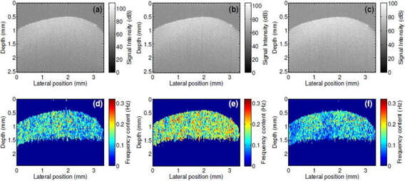
 Most of the fundamental pathological processes in living tissues exhibit changes at the nanoscale. Noninvasive, label-free detection of structural changes in biological samples pose a significant challenge to both researchers and healthcare professionals. Modern nanoscopy largely requires labeling, is limited to superficial 2D imaging, and is generally not suitable for in vivo applications. In this article published in Applied Physics Letters, we presented a method, spatial frequency domain correlation mapping optical coherence tomography (sf-cmOCT), developed at NUI Galway, for detection of depth resolved nanoscale structural changes noninvasively. Our approach is based on detection and correlation of the depth resolved spectra of axial spatial frequencies of the object which are extremely sensitive to structural alterations. These experimental results demonstrate possibilities for the study of nanoscale structural changes, without the need for biomarkers or labels. Thus, sf-cmOCT offers exciting and far-reaching opportunities for early disease diagnosis and treatment response monitoring, as well as a myriad of applications for researchers. Full reference: Sergey Alexandrov, Paul M. McNamara, Nandan Das, Yi Zhou, Gillian Lynch, Josh Hogan, and Martin Leahy. Spatial frequency domain correlation mapping optical coherence tomography for nanoscale structural characterization. Applied Physics Letters 115:12 (2019). Full article here 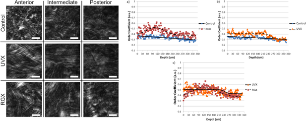 One option for treating keratoconus, a degenerative disease that results in a weakened cornea and vision loss, is cross-linking. Cross-linking strengthens the overall structure of the cornea by promoting the creation of covalent bonds between neighboring collagen fibrils or between fibrils and the surrounding extracellular matrix. To measure how cross-linking affects the cornea at different depths inside the stroma, three different corneal treatments were performed on rabbit animal models: no treatment, Riboflavin instillation/Ultraviolet irradiation, and Rose Bengal instillation/Green light irradiation. Rabbits were sacrificed one and two months after treatment. Images of corneal lamellae were taken with a second harmonic generation laser scanning microscopic developed at VioBio Lab (CSIC) and were analyzed with a MATLAB script to measure the different directions of the collagen fibers. The collagen fibers in the cross-linked corneas had more uniform fiber orientations than in untreated corneas through a depth of 300 microns. The amount of second harmonic generation is also greater in cross-linked corneas than in control corneas. Both the organization and second harmonic generation signal increased between one month and two months after treatment. This research was a collaboration between CSIC, the University of Valladolid, and the Wellman Center for Photomedicine.
Full reference: James A. Germann, Eduardo Martínez-Enríquez, M. Carmen Martínez-García, Irene E. Kochevar, Susana Marcos; Corneal Collagen Ordering After In Vivo Rose Bengal and Riboflavin Cross-Linking. Invest. Ophthalmol. Vis. Sci. 61(3):28 (2020). Full article here 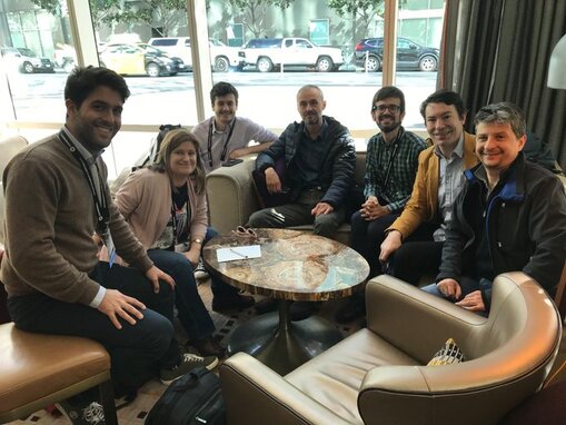 Imcustomeye partners were present at the SPIE Photonics West that took place in San Francisco this February 2020. SPIE Photonics West is the premier laser, photonics, biomedical optics event, organized by The International Society for Optics and Photonic (SPIE). The not-for-profit society promotes emerging technologies through interdisciplinary information exchange, continuing education, publications, patent precedent, and career and professional growth.
A progress meeting of the Imcustomeye project was held the past February 5 coinciding with the presence of CSIC, NUIG, PAN and 2EyesVision partners at the Conference. The meeting covered a wide range of topics including status of the technology, prospects for clinical measurements and project communications. Here are the Imcustomeye contributions at the SPIE Photonics West: Sunday 2 February - Ocular Biomechanics Session. 9:40 am Susana Marcos “Corneal dynamic imaging and second harmonic generation to evaluate in vivo corneal cross-linking” (Invited Paper). Consejo Superior de Investigaciones Científicas (Spain). - Ocular Biomechanics Session. 9:10am Andrea Curatolo “Customized swept-source optical coherence tomography system for air-puff induced corneal deformation imaging on multiple meridians” A. Curatolo, J. Birkenfeld, E. Martínez, J.A. Germann, D. Pascual, G. Muralidharan, Consejo Superior de Investigaciones Científicas (Spain); J. Palací, 2Eyes Vision SL (Spain); J. Solarski, K. Karnowski, M. Wojtkowski, Institute of Physical Chemistry (Poland); S. Marcos, Consejo Superior de Investigaciones Científicas (Spain). - Poster Session. Simultaneous optical coherence tomography measurements at two arbitrary meridians, Karol Karnowski, J. Solarski, A. Consejo, M. Wojtkowski, Institute of Physical Chemistry (Poland). - Poster Session. Light intensity distribution from corneal Scheimpflug images as a predictor of eye diseases, Alejandra Consejo, Polish Academy of Sciences (Poland); K. Karnowski, J. Solarski, Institute of Physical Chemistry PAS (Poland); D.R. Iskander, Wroclaw Univ. of Science and Technology (Poland); M. Wojtkowski, Institute of Physical Chemistry PAS (Poland). - Poster Session. Use of optical coherence yomography to access corneal vibrational resonance. Ryan McAuley, A. Nolan, S.A. Alexandrov, M.J. Leahy. National Univ. of Ireland, Galway (Ireland). |
 RSS Feed